
Expertise
Histology platform
> Materials and Techniques
Frozen tissue section
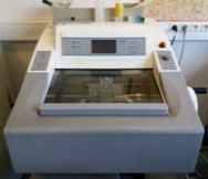
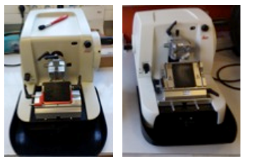
N°1: cryostat Microm HM560manual and automatic.
Knife and specimen holder temperature independent, with suction.
Description : Equipment that allows the production of frozen sections from 5 µm to 30 µm, from fresh or fixed material with the maintain of low temperatures ranging from 10°C to -50°C. All types of previously prepared samples can be cut using these cryostats.
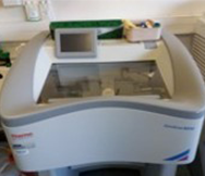
N°2: Cryostat ThermoScientific Cryostar NX50 manual.
Adjustable specimen temperature, suction and decontamination of the work chamber by vaporization.
Description : Equipment that allows the production of frozen sections from 5 µm to 30 µm, from fresh or fixed material with the maintain of low temperatures ranging from 10°C to -50°C. All types of previously prepared samples can be cut using these cryostats.
Paraffin embedded tissue section
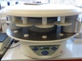
Impregnation machine Myr STP120
Description : Equipment that allows the progressive dehydration of the fixed samples through increasing baths of ethanol, Histoclear (substituting for xylene) and ending by impregnation in liquid heated paraffin.
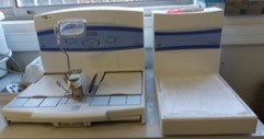
Paraffin embedding station Myr EC350
Description : : Embedding station composed of 2 modules: a heating paraffin distributor and a cryo-console. After impregnation in paraffin, the samples are embedded in a paraffin block.
Leica MR2125RTS and MR2125RT
Description: Microtomes allow the production of thin sections from 4 µm to 40 µm from paraffin blocks.
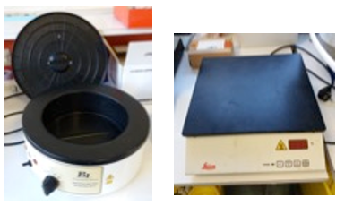
Barnstead Water Bath and Leica HI1220 Hot Plate
Description: The bath allows to recover the sections of the blocks and to collect on specific glass slides (Super Frost or Polysin). The heating plate allows flattening adhesion of the section on slides.
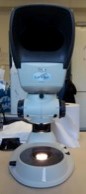
Lynx Vision engineering magnifier
Description: Table magnifier for observing samples at high magnification before and after coloring.
> Applications and samples evaluated
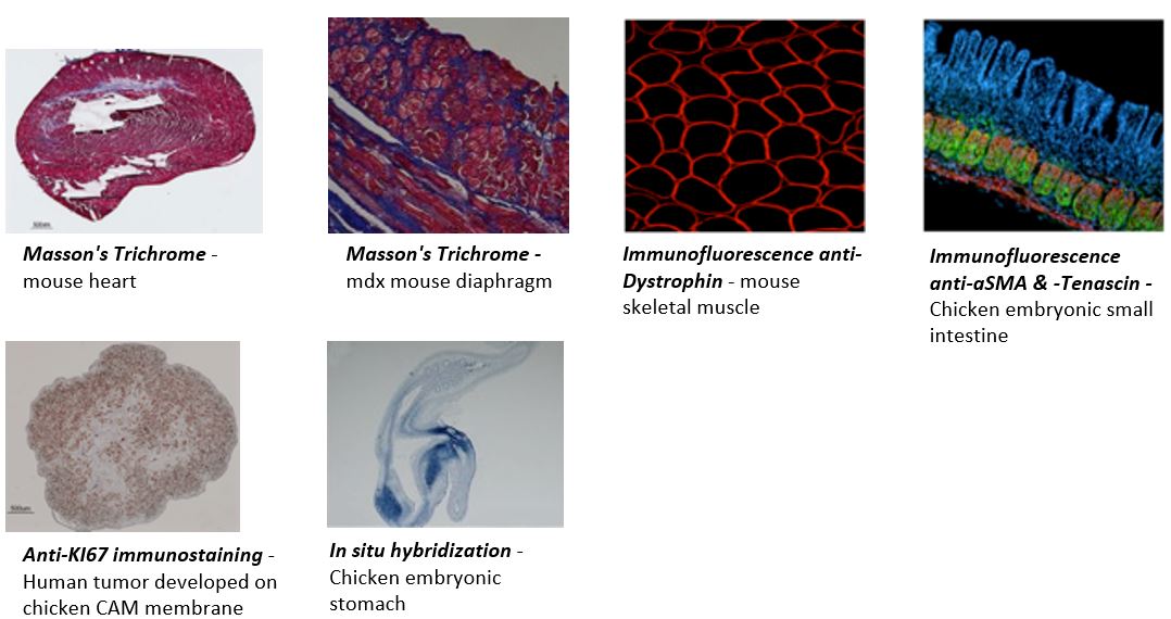
Description: Applications in optical microscopy can be diverse, ranging from simple histo-morphological staining, to immuno-labeling of proteins with a choice of varied detection: fluorochromes or enzymatic activities (HRP, AP, etc.) and the detection of specific mRNA by the technique of in situ hybridizatio
Tissues evaluated: Heart; Skeletal muscle; Gastrointestinal tube; Lung.
Models assessed: Human; Mouse; Rat; Chicken.